


Bacteria - cowpea

Contributors to this section: IITA, Nigeria (M. Ayodele, L. Kumar).
|
Contents: |
Cowpea bacterial blight, Bacterial blight, Leaf spot
Scientific name
Xanthomonas axonopodis pv. vignicola (Burkholder 1944) Vauterin et al. 1995
Other scientific names
Xanthomonas phaseoli f.sp. vignicola (Burkholder) Sabet 1959
Xanthomonas campestris pv. vignicola (Burkholder 1944) Dye 1978
Importance
High
Significance
Although yield losses from the fields have been reported, they have not been quantified
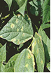
Symptoms
Leaves: pin point water soaked spots on the leaves.
Spots coalesce to form orange lesions surrounded by yellow halo. The bacteria infects also the stems, causing cracking, canker and the pods causing water soaked spots.
Hosts
The major hosts of the bacterial blight pathogen are Vigna unguiculata (cowpea), Crotalaria juncea (sunn hemp), Lablab purpureus (hyacinth bean), Phaseolus vulgaris (common bean), Solanum nigrum (black nightshade), Tephrosia purpurea (purple tephrosia), Vigna mungo (black gram).
Geographic distribution
China, India, Turkey, Botswana, Egypt, Nigeria, S Africa, Sudan, Tanzania, Zimbabwe, Puerto Rico, USA.
Biology and transmission
The bacterium is gram- negative rod, single or in pairs motile by one polar flagellum.Colonies on NBY are yellow and circular. Two biotypes have been isolated and identified morphologically from infected cowpea fields in Nigeria. Isolate 1 which produces yellow colonies on NBYis not sensitive to antibiotic lincomycin while isolate 2 which produces dark yellow/light brown colonies on NBY media is sensitive to lincomycin.
The pathogen is seed borne and seed transmitted. Disease development and spread is favoured by rainfall and the bacterium survives in crop residues.
Detection/indexing methods
Agar method and Serology
Agar test using NBY media, selective media and biochemical analysis:
- Randomly select a subsample of 500 seeds (or less if fewer seeds are available) .
- Surface-sterilise the seeds by placing them in a 10% sodium hypochlorite solution for 3 minutes.
- Rinse the seeds with sterile distilled water, blot dry on sterile paper towel, and place seeds equidistantly on NBY agar media in a 9-cm petri dish.
- Incubate seeds at 25oC under 12 h fluorescent light or 12 h NUV light for 4 days.
- Inspect each seed carefully under the stereomicroscope. Use a needle or forceps to turn the seed over and to examine the under side.
- Use the compound microscope for the identification of the bacterium
- Pick bacteria colony and streak unto new NBY plates
- Incubate at 28oC for 48hrs
- On a clean slide, thinly spread bacterial colony; gram stain
- Subculture selected bacteria for further identification on NBY.
- Subject the bacteria to biochemical tests.
- Spot plate the bacterium on MSP and M71 selective media for specie confirmation.
Serology: ELISA using polyclonal antibody for the detection of the bacterium The polyclonal antibody for the detection of X. axonopodis pv vignicola is available in IITA.
Treatment/control
Screen house/ containment facility inspection in collaboration with the National Plant Quarantine service Inspectors for certification and issuance of phytosanitary certificates.
- Seed for export grown in the screen house
- Inspection during active growth of all the multiplication sites
- Use of resistant varieties
- Seeds for international distribution grown under seed certification schemes and Pest Free areas
Procedures in case of positive test
Discard
For import: Grow seeds under containment. Inspect during active growth.Laboratory testing of vegetative parts (leaves and stem ), seeds using agar method and ELISA
Positive lines discard, not acceptable for international distribution in compliance to National Plant Protection Organization requiring additional declaration of freedom from the X. axonopodis pv vignicola or that X. axonopodis pv vignicola is not known to occur in the country of origin or multiplication sites.
References and further reading
Elliot C. 1951. Manual of bacterial plant pathogens. 2d ed Chronica Botanica:Waltham, Mass
Moretti C, Mondjana AM, Zazzerini A, Buonaurio R. 2006. Occurence of leaf spot on cowpea (Vigna unguiculata) caused by Xanthomanas axonopodis pv. vignicola in Mozambique. [online].
Patel PN, Jindal JK. 1970. Indian Phytopath. Soc. Bull. 6, 28-34.
Watkins GM. 1943. Pl. Reptr. 27, 556
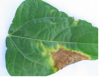
Field symptom (photo: IITA) |
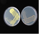
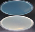
yellow and pale yellow colonies (photo: IITA) |
Field symptom (photo: IITA) |
Scientific name
Xanthomonas axonopodis pv. vignae
Other scientific names
Not reported.
Importance
High
Significance
Not reported.
Symptoms
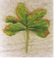
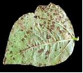
It is a foliar disease. The symptoms are visible on the leaves. On the leaves, the symptoms start as small tiny water soaked dots on the underside of the leaves. The dots coalesce to form circular, raised dark water soaked spots on the underside of the leaves and dark brown necrotic spots on the upper side of the leaves’ Older lesions/ pustules become sunkened and dried in the center.
Leaves turn yellow under severe infection. Defoliation is also a symptom on plants with severe infection. In the field, there is usually some confusion between rust infection (fungal pustules) and bacterial pustules. The first field difference between the two pathogens, is the presence of brown/ pink dust from the leaves of the infected cowpea plants during active growth, this indicates that the pathogen in question is the rust fungi and not a bacterium.
 Bacterial pustule (photo:IITA) |
Hosts
Cowpea.
Geographic distribution
Widely spread in the humid and savannah eco regions where cowpea is grown in Nigeria. The disease has also been reported in Tanzania and Brazil.
Biology and transmission
The pathogen is seed borne and seed transmitted. Disease spread is favoured by rains
Detection/indexing methods used at IITA
Agar method, Serology and leaf isolation:
Agar test using NBY media, selective media and biochemical analysis:
- Randomly select a subsample of 500 seeds (or less if fewer seeds are available) .
- Surface-sterilise the seeds by placing them in a 10% sodium hypochlorite solution for 3 minutes.
- Rinse the seeds with sterile distilled water, blot dry on sterile paper towel, and place seeds equidistantly on NBY agar media in a 9-cm petri dish.
- Incubate seeds at 25oC under 12 h fluorescent light or 12 h NUV light for 4 days.
- Inspect each seed carefully under the stereomicroscope. Use a needle or forceps to turn the seed over and to examine the under side.
- Use the compound microscope for closer for the identification of the bacterium
- Pick bacteria colony and streak unto new NBY plates
- Incubate at 28oC for 48hrs
- On a clean slide, thinly spread bacterial colony; gram stain
- Subculture selected bacteria for further identification on NBY.
- Subject the bacteria to biochemical tests.
- Spot plate the bacterium on MSP and M71 selective media for species confirmation.
Serology:
Conduct ELISA using polyclonal antibodies for the detection of the bacterium.
Treatment/control
- Seed for export grown in the screen house
- Inspection during active growth
- Use of resistant varieties
- Seeds for international distribution grown under seed certification schemes and Pest Free areas.
Procedures in case of positive test
Discard.
At IITA, for import: Grow seeds under containment. Inspect during active growth. Laboratory testing of vegetative parts (leaves and stem ), seeds using agar method and ELISA.
Positive lines discard, not acceptable for international distribution in compliance to National Plant Protection Organization requiring additional declaration of freedom from X. axonopodis pv vignae or X. axonopodis pv vignae not known to occur in the country of origin or multiplication sites.
References
Patel PN. 1978. 3rd Int. Congr. Pl. Path, Munich, August, 1978. p 72 ( abstr.).
Williams RJ. 1975. PANS. 21, 253-267.
Bacterial halo blight, Halo blight (of beans), Grease spot (of beans), Bacterial bean blight
Scientific name
Pseudmonas syringae pv phaseolicola.
Other scientific names
Pseudomonas medicaginis f.sp. phaseolicola (Burkholder) Dowson 1957
Pseudomonas vignae Gardner & Kendrick
Importance
High
Significance
Halo blight is world wide in distribution in the bean growing regions. Walker and Patel, (1964), reported that epidemics have been recorded in some parts of the USA
In some trials with artificial inoculations, losses in seed yield ranging from 2.8-55.4% were obtained (Anon., 1980). Yield losses of 43% in the UK and between 23 and 43% in Michigan were reported by Allen et al. (1998).
Allen et al. (1998) observed crop losses as a result of halo blight in Lesotho, Rwanda and Zimbabwe.
Symptoms
The pathogen infects all the growing stages of the plant: flowering, podding , pre-emergence, and seedling. The pods, growing points, leaves, seeds, stems and whole plant are also infected.
Symptoms found on the various plant parts are :
Leaves: water-soaked spots that later turn red-brown and necrotic. Lesions; abnormal colours; lime-green halo around the necrotic lesion. Pod: water-soaked, greasy spots that vary in size with brown margins. Seeds: rot, shrivelling and discoloured, some times infected seeds are symptomless. Stem: girdling and rotting of nodes, discoloration; and exudates. Whole plant: seedling blight, chlorosis, dieback, lime green coloration, stunting and distorted (Allen et al., 1998).
Hosts
Cowpea (Vigna unguiculata) is considered to be one of the minor host of the bacterium.
The major hosts infected by the bacterium are: Phaseolus acutifolius (tepary bean), Phaseolus coccineus (runner bean), Phaseolus lunatus (lima bean), Phaseolus vulgaris (common bean).
The minor hosts are: Cajanus cajan (pigeon pea), Centrosema , Desmodium (tick clovers), Glycine max (soyabean), Lablab purpureus (hyacinth bean), Pisum sativum (pea), Pueraria montana var. lobata (kudzu), Vigna angularis (adzuki bean),and Vigna radiata (mung bean).
Geographic distribution
Worldwide
Biology and transmission
Non-sporulating, Gram-negative, aerobic rods. Motile by means of multitrichous polar flagellae. Bacterial colonies are white to cream on agar medium with a bluish colour, producing a green fluorescent pigment on King's medium B agar. Optimum growth temperatures for the bacterium are 20-23°C.
Nine races of the halo blight pathogen isolated from Africa and other bean growing areas, have been characterized based on their reactions to eight differential cultivars. (Taylor et al., 1996). Ariyarathne (1997) identified two new races that occurred in Nebraska, USA.
The pathogen can be stored for up to 5 years at -20°C (Schwartz, 1989).
The disease is seed borne and seed transmitted.
The pathogen survives in infected seed and plant residues on the soil surface. Halo blight is favoured by cool, wet weather (Allen et al., 1996). The bacteria multiply rapidly under favourable environmental conditions with or without the formation of lesions. The water-soaking results from extracellular polysaccharides from bacterial slime interacting with plant tissue (El-Banoby and Rudolph, 1979 After penetration, symptoms develop within 6-10 days at 24-28°C. Halo formation is more common at 16-20°C. Lesions can be without halo at temperatures of 28°C and above (Schwartz, 1989). The bacterium produces a toxin, phaseolotoxin, which contains N-phosphosulfamylornithine. This toxin is responsible for the typical halo symptoms and general chlorosis (Schwartz, 1989).
Detection/indexing methods used at IITA
Agar test using NBY media:
- Randomly select a subsample of 500 seeds (or less if fewer seeds are available).
- Surface-sterilise the seeds by placing them in a 10% sodium hypochlorite solution for 3 minutes.
- Rinse the seeds with sterile distilled water, blot dry on sterile paper towel, and place seeds equidistantly on NBY agar media in a 9-cm petri dish.
- Incubate seeds at 25oC under 12 h fluorescent light or 12 h NUV light for 4 days.
- Inspect each seed carefully under the stereomicroscope. Use a needle or forceps to turn the seed over and to examine the under side.
- Use the compound microscope for closer for the identification of the bacterium
- Pick bacteria colony and streak unto new NBY plates
- Incubate at 28oC for 48hrs
- On a clean slide, thinly spread bacterial colony; gram stain
- Subculture selected bacteria for further identification on NBY.
- Subject the bacteria to biochemical tests.
- Spot plate the bacterium on MSP and M71 selective media for specie confirmation.
Treatment/control
- Removal of infected debris after harvest
- Crop rotation using cereals
- Plant disease free, certified healthy seeds and resistant varieties
- Production of seeds under certification scheme and established Pest Free area
- Field inspection during active growth
- Post entry quarantine processing: ELISA , and growing on test under containment to prevent export/ import of infected seeds for research and conservation
Procedures in case of positive test
Discard
For import:
- Post entry quarantine processing by growing the lines under containment facility to intercept the bacteria.
- Inspection during active growth in company of the Plant quarantine inspectors
- Rogue lines with symptoms and incinerate
- ELISA testing for all symptomless lines
- Discard positive lines by incineration
For export:
- Seeds harvested from the multiplication fields are seed health tested
- Using the agar method and selective media previously described in addition to ELISA test. Infect lines are discarded by incineration.
- Not acceptable for international distribution
References and further reading
Allen DJ, Buruchara RA, Smithson JB. 1998. Diseases of common bean. In: Allen DJ, Lenne J, editors. The Pathology of Food and Pasture Legumes. Wallingford, UK: CAB International, 214.
Anon. 1980. Germplasm screening for desirable variability. Disease loss studies. Centro Internacional de Agricultura Tropical: 1979 Bean program. Annual Report.1980, 19-22.
Ariyarathne HM. 1997. Pathogenic variation for the halo blight bacterium and mapping of loci for multiple diseases in common bean. PhD diss. University of Nebraska, Lincoln
El-Banoby FE, Rudolph K. 1979. A polysaccharide from liquid cultures of Pseudomonas phaseolicola which specifically induces water-soaking in bean leaves (Phaseolus vulgaris L.). Phytopathologische Zeitschrift, 95(1):38-50;
Schwartz HF. 1989. Halo blight. In: Schwartz HF, Pastor-Coralles MA, editors. Bean Production problems in the Tropics. Cali, Colombia: Centro Internacional de Agricultura Tropical (CIAT).
Taylor JD, Teverson DM, Allen DJ, Pastor Corrales MA. 1996. Identification and origin of races of Pseudomonas syringae pv. phaseolicola from Africa and other bean growing areas. Plant Pathology
Walker JC, Patel PN. 1964. Splash dispersal and wind as factors in epidemiology of halo blight of bean. Phytopathology, 54:140-141.
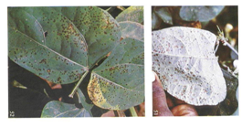
Field Symptom (photo: IITA) |
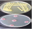
Growth on MSP (photo: IITA) |
Control plate of MSP (photo: IITA) |
Scientific name
Pseudmonas syringae pv syringae
Other scientific names
Phytomonas syringae (van Hall) Bergey et al. 1930
Pseudomonas vignae var. leguminophila (Burkholder) Magrou & Prévot 1948
Importance
High
Significance
P. syringae pv. syringae is seedborne on several crops throughout the world. It attacks several economic crops causing great losses. The bacterium was reported to have caused epidemics on Phaseolus vulgaris cvBonusplantingsin the Transvaal highveld in South Africa where crop losses of up to 55% were reported. The pathogen was also detected in commercial seed stocks (Serfontein, 1994).
Symptoms
-
P. syringae pv. syringae attacks the flowering, podding , post-harvest, seedling and vegetative growing stages of the plant. It also affects the leaves, stems, inflorescence, pods, seeds, roots, and the whole plant
-
Some infected plants are symptomless
-
The bacterium causes frost injury to plants, at relatively high freezing temperatures
-
Symptoms found on the various plant parts are:
-
Leaves : small, water-soaked spots first appearing on the lower sides of the leaves, the spots enlarge, coalesce, form necrotic lesions, blacken and die
-
Stem: water-soaked, sunken brown lesions, splitting at the surface, girdling,
-
Pods: small water-soaked spots , enlarge, coalesce, turn brownish or reddish colored with age (Agrios, 1988; Hall, 1991).
-
Seeds: discoloration, spots, and
-
Seedling: dieback.
 Field symptom (photo:IITA) |
Hosts
The pathogen has several hosts comprising of mono and dicots.
The major hosts of Ps syringae pv syringae are : Vigna unguiculata , (cowpea)Vitis , (grape)Vitis vinifera , (grapevine)Zea mays (maize)Abelmoschus esculentus , (okra)Allium cepa , (onion)Allium porrum , , (leek)Citrus aurantium , (sour orange)Citrus limon , (lemon)Citrus maxima , (pummelo)Citrus medica , (citron)Citrus reticulata , (mandarin)Citrus sinensis , (navel orange)Citrus x paradisi , (grapefruit)Coffea arabica , (arabica coffee)Cucumis sativus , (cucumber)Cucurbita , (pumpkin)Cucurbita maxima , (giant pumpkin)Cyphomandra betacea , (tree tomato)Juglans regia , (walnut)Lablab purpureus , (hyacinth bean)Lactuca sativa , (lettuce)Lycopersicon esculentum , , (tomato)Malus domestica , (apple)Mangifera indica , (mango)Medicago sativa , (lucerne)Musa x paradisiaca , (plantain)Nicotiana tabacum , (tobacco)Oryza sativa , (rice)Panicum , (millets)Panicum miliaceum , (millet)Passiflora edulis , (passionfruit)Pennisetum glaucum , (pearl millet)Pennisetum purpureum , (elephant grass)Persea americana , (avocado)Phaseolus coccineus , (runner bean)Phaseolus lunatus , (lima bean)Phaseolus vulgaris , (common bean)Piper nigrum , (black pepper)Pisum sativum , (pea)Prunus amygdalus , Prunus armeniaca , (apricot)Prunus avium , (sweet cherry)Prunus domestica , (plum)Rosa , (roses)Sorghum bicolor , (sorghum)Sorghum halepense , (Johnson grass)Sorghum sudanense , (Sudan grass)Triticum aestivum , (wheat)Vicia faba , (broad bean)Vicia villosa , Vigna angularis (adzuki bean)
The only recorded minor host is Chenopodium quinoa (quinoa)
Geographic distribution
Worldwide.
Biology and transmission
P. syringae pv. syringae is an aerobic, unicellular Gram-negative rod, motile having one to several polar flagella. The bacterial colonies are circular, milky-white, raised, glistening, translucent, smooth surface, and entire margin. It produces a green fluorescent pigment on King's B medium. The bacteriium produces two lipopeptide toxins, syringomycin and syringopeptin. (Hutchison and Gross, 1997.
This species is represented by strains which are heterogeneous genetically (Gardan et al., 1997). P. syringae pv. syringae comprise of more than 50 distinct pathogens identified as pathovars (Dye et al., 1980; Young et al., 1996). P. syringae pv. syringae is a pathovar originally isolated from lilac but now found infecting several hosts. The pathogen survives on a number of crops and non-crop species, which serve as sources of primary inoculum for infection ( Hall, 1991a, b.). The bacterium is found in the soil, water and on plant surfaces. The bacterium is seed borne and seed transmitted. Spreads through plant parts, rain and wind.
On cowpea, P. syringae pv. syringae, survives in infected seeds and stems. From the seed, it infects the cotyledons, spread to the leaves or enter the vascular system and cause systemic infection resulting in stem and leaf lesions. Disease spread and intensity is favored by rains.
Detection/indexing methods at IITA
Agar method NBY, followed by:
- Selective medium
- and Semi-selective medium
- Serology ( ELISA)
Treatment/control
Seed treatment using mancozeb has not been effective.
- Use of disease-free seeds
- Plant resistant varieties. These are available in IITA
- Crop rotation, discard of plant debris after harvest
- Seed multiplication for international distribution in Pest Free Areas.
Procedures in case of positive test
For import:
- Post entry quarantine processing by growing the lines under containment facility to intercept the bacteria.
- Inspection during active growth in company of the plant quarantine inspectors
- Rogue lines with symptoms and incinerate
- ELISA testing for all symptomless lines
- Discard positive lines by incineration
For export:
- Seeds harvested from the multiplication fields are seed health tested
- Using the agar method and selective media previously described in addition to ELISA test. Infected lines are discarded by incineration.
- Not acceptable for international distribution
References and further reading
Agrios GN. 1988. Plant pathology. London, UK: Academic Press Inc. (London) Ltd.
CAB International. 2007. Crop Protection Compendium, 2007 Edition. Wallingford, UK: CAB International
Goszczynska T, Serfontein JJ. 1998. Milk-Tween agar, a semiselective medium for isolation and differentiation of Pseudomonas syringae pv. syringae, Pseudomonas syringae pv. phaseolicola and Xanthomonas axonopodis pv. phaseoli. Journal of Microbiological Methods, 32(1):65-72.
Hall R. 1991. Compendium of Bean Diseases. St Paul, Minnesota, USA: APS Press.
Higley PM, McGee DC, Burris JS. 1993. Development of methodology for non-destructive assay of bacteria, fungi and viruses in seeds of large-seeded field crops. Seed Science and Technology, 21(2):399-409
Hutchison ML, Gross DC. 1997. Lipopeptide phytotoxins produced by Pseudomonas syringae pv. syringae: comparison of the biosurfactant and ion channel-forming activities of syringopeptin and syringomycin. Molecular Plant-Microbe Interactions, 10(3):347-354
Mohan SK, Schaad NW. 1987. An improved agar plating assay for detecting Pseudomonas syringae pv. syringae and P. s. pv. phaseolicola in contaminated bean seed. Phytopathology, 77(10):1390-1395
Serfontein JJ. 1994. Occurrence of bacterial brown spot of dry beans in the Transvaal province of South Africa. Plant Pathology, 43(3):597-599.
Comments
- No comments found
Leave your comments
Post comment as a guest