


Establishment of aseptic cultures of banana

|
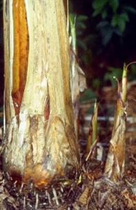
Mother plant and young shoots – source of material (photo: IITA)
|
Contributors to this page: Bioversity International, Belgium (Ines Van den Houwe); IITA, Nigeria (Dominique Dumet, Badara Gueye).
New accessions can be received as small samples of in vitro cultures from other collections, seeds of wild types or as classical vegetative propagation materials (apex-containing parts of the banana plant). When materials come from the field, a series of procedures are required and are described below:
Initiation of shoot tip cultures from suckers
In Musa, explants can be obtained from all plant parts that contain a shoot meristem: the parental pseudo stem, its suckers, peepers, sword suckers, lateral buds or very small eyes.
- Explants should preferably be isolated from young, vigorous and healthy looking suckers of 40-100 cm in height with a corm diameter of about 10 cm.
- These should be collected from acharacterized flowering mother plant to guarantee trueness-to-type.
Usually only the apical meristem is available from a trimmed sucker. Eventually, smaller buds (sleeping eyes) on the corm can also be used as explants for tissue culture initiation.
Explant isolation and disinfection
- Excise from the sucker a cubical block of tissue (2x2x2 cm) containing the apical meristem.
- If there are buds on the corm surface present, excise the buds surrounded by 0.5 cm of corm tissue.
Surface sterilization of explants
- Transfer each cube of tissue to a different Erlenmeyer flask.
- Soak each individual cube of tissue in ethanol (96%) and shake gently.
- Immediately decant and rinse for 30 seconds in sterile deionised water.
- Transfer the Erlenmeyer flask to a laminar air flow cabinet.
- Emerse each plant tissue cube for 20 minutes in a 2% hypochlorite solution or diluted commercial bleach solution supplemented with a few drops of Tween 20 (surfactans). Liquid dish-washing detergent can be used if no Tween is available.
- Cover the Erlenmeyer flasks with a glass petri dish lid and swirl the solution frequently.
- Decant the hypochlorite solution.
- Then rinse three times in the space of ten minutes with sterile deionised water and decant.
Excision of explants
Shoot apices of bananas are enclosed in many tightly overlapping leaf initials. The shoot tip is about 2-5 mm in size and consists of the meristem covered with several leaf primordia supported on a small base of corm tissue.
- Remove the outer tissue that was exposed to the disinfectant solution from all sides of the cube.
- Sterilize dissection instruments repeatedly during manipulation by flame of glass bead sterilizer.
- Remove stepwise the covering leaf primordia, cut away as much corm tissue as possible, as this causes blackening of the culture during initiation.
In case of high risk of bacterial contamination, i.e. when suckers are harvested during the wet season, it is recommended to excise a smaller explant consisting of the meristem covered with 1-2 leaf primordia (1 mm).
- To excise the meristem tip, place the excised shoot tip under a dissecting microscope (working in a laminar flow cabinet) and further reduce the size by removing leaf primordia and some corm tissue at the base of the explant.
Tissue culture initiation
- Immediately place the excised explant onto culture initiation medium.
- Push the explant slightly into the culture medium to ensure good contact.
- Keep the cultures in complete darkness for one week to reduce blackening (photo-oxidation of polyphenols).
- Growth of the tissue should start about two weeks after inoculation, accompanied by a swelling of the leaf sheet tissue and corm tissue, a change of colour from white to green, and abundant oxidation of polyphenolic compounds that are released from the corm tissue (blackening of the tissue and surrounding growth medium).
- Transfer the explants to fresh medium every two weeks during the first eight weeks in culture.
- Trim the blackened and/or swollen corm tissue, if necessary, leaving the viable leaf primordia tissue of the explant intact.
- After 10-12 weeks the explant will develop into a shoot.
- Record the survival rate and visible contamination at each transfer for each replicate.
The size of the explant is an important factor in the successful culturing of shoot tips of bananas. Very small explants increase the chance of producing bacteria-free and virus-free cultures, but the mortality rate is high. Intermediate sized explants produce clean vigorous cultures that multiply rapidly whereas very large explants tend to show more blackening and contamination, and thus lower rates of successful tissue culturing.
Initiation of tissue cultures from seeds
For wild banana species, seeds can also be used to initiate tissue cultures, if no vegetative propagation material is available. However germination of banana seeds is known to be low and erratic, therefore preference is given to direct culturing of the axenic embryo isolated from the seed (= embryo rescue).
Surface disinfection of banana seeds
- Soak the seeds for 2-3 days in tap water, without stirring, in an Erlenmeyer flask. Refresh the water daily in order to avoid fungal contamination or growth.
- Soak the seeds for two minutes in EtOH (95 % v/v) in an Erlenmeyer flask and cover the flasks with a lid.
- Decant and transfer the flasks to a laminar air flow cabinet. Disinfect the seeds in a 1.5% hypochlorite solution and 1-2 drops of Tween for 20 minutes and shake gently. Wash the sterilized seeds with sterile deionised water rinsing twice in 15 minutes.
Embryo isolation
- Use forceps to fix the seed on a sterile cutting pad.
- Cut the seed lengthwise next to the micropyle.
- Remove the plug and pick up the embryo.
- Place the embryos on embryo germination medium in glass test tubes.
- Incubate the embryos under normal conditions for banana shoot tip cultures.
Germination can start about two days after inoculation, accompanied by a colour change from white to yellow and abundant formation of root hairs. Once cultures are successfully initiated, regular testing of the plant material for asepsis should start.
Contamination indexing (bacteria)
A primary requirement in the establishment and permanent maintenance of an in vitro collection is the asepsis of the tissue cultures. Sterility of the cultures is also of paramount importance for their intercontinental exchange and whenever cultures are used for various research purposes.
To safeguard the in vitro cultures against microbial contamination, and to prevent the spread of contaminants in the tissue culture system, monitoring of the material for asepsis should be performed at crucial steps (see Tissue culture testing below) and at regular intervals during the processing of material in the genebank.
Visual examination
- Bacterial contamination can be detected as faint ‘clouds’ in the medium around the basal part of the explants or culture. Therefore, the use of a clear gelling agent (Gelrite) in tissue culture will facilitate the detection of contamination.
- Sometimes even colony growth is detected around the tissue on the surface of the growth medium.
Most of the time however the contaminants remain deep seated inside the plant tissue and only appear in the growth medium under favourable conditions (increased sucrose concentration, change of pH of the medium, increase of the ambient temperature, deterioration of the plant tissue etc.).
Tissue culture testing
Tissue culture testing should usually be done upon introduction of new material, during storage and at annual subculturing. The screening test is simple and non-destructive. Testing involves streaking of the basal part of the shoot tip onto a semi-solid bacteriological medium.
- The test should be done at subculturing just before transferring the explant to the growth medium.
- A single shoot tip should be isolated from the culture and trimmed to a suitable size for transfer to fresh culture medium.
- The corm basis of this shoot tip should be streaked three times onto bacteriological medium and immediately placed onto plant growth medium.
- The petri dishes should be sealed with parafilm and incubated at 28°C.
- The standard testing medium is a broad spectrum bacteriological medium, which allows growth of a wide range of bacteria. The medium includes nutrient agar (8%), enriched with yeast extract (5 g/l) and glucose (10 g/l).
- Most bacteria are fast growing and will appear on the medium after two or three days, but slow growing contaminants may require up to eight weeks to appear on the medium. It is thus important to observe the medium for at least two months after testing.
-
In case of inconclusive results, alternative and complementary detection methods can be applied:
- Using specific/selective growth medium.
- Macerating the tissue and incubating in liquid the bacterial growth medium.
- Using polymerase chain reaction (PCR) methods.
Decontamination treatment
Contaminated cultures should be eliminated by autoclaving. However, when all replicates of one accession at initiation or in storage are tested positive, contaminated material cannot simply be discarded as the accessions may be unique and difficult to replace. They should then be subjected to a decontamination treatment.
- Before applying a decontamination treatment it is recommended to characterize the isolated bacteria in order to determine the most effective method of eradication.
- For routine application, meristem culture, for which only a rough characterization of the contaminant is required, is preferred over treatments with antibiotics of which the therapeutic success depends on the identification of the contaminant, bacterial sensitivity and phytotoxicity. Moreover, the application of antibiotic agents may involve the risk of developing resistant bacterial strains.
Elimination of bacteria through meristem culture
This technique involves the isolation of aseptic meristems from the contaminated in vitro cultures. Depending on the type of contaminating bacteria, the eradication method is further refined by subjecting either contaminated in vitro or greenhouse grown contaminated plants to meristem culture.
- Culturing 1 mm sized meristems, directly isolated from contaminated in vitro plants, is found most effective to eliminate bacteria like Bacillus sp.
- The shoot tip must be isolated from the contaminated culture and further reduced to 1 mm in length under a dissecting microscope. Young leaves must be removed until the dome covered by two to three leaf primordial remains.
- It is important that the corm tissue is carefully and maximally removed, as bacteria often reside in this tissue.
- The excision of the meristem tip should be done under stringent aseptic conditions in order to minimize the risk of transferring bacteria to the aseptic inner meristematic zone of the explant.
- It is essential that the explant material only comes into contact with sterile instruments and surfaces. Between use, forceps and scalpels should be sterilized (in a glass bead sterilizer or flame) and the cutting pad should be sterilized before the surface comes into contact with a freshly cut tip.
Elimination of bacteria from greenhouse plants
In some cases, for instance when the tissue cultures are contaminated with very small sized bacteria, the method described above is less effective. For a successful elimination of these bacteria, the explant size might need to be further reduced. These smaller sized explants, however, often fail to establish in vitro. Therefore, an alternative approach to eliminate bacteria is to isolate explants from greenhouse plants, acclimatizing the in vitro plants.
- The contaminated in vitro plants should be transferred to potting soil in PET pots in a greenhouse and grown for a period of 3-6 months or until the stems reach a diameter of at least 5 cm.
- The plants should be kept dry for 2-3 weeks and then explants of 1-3 mm tips can be isolated to re-initiate in vitro cultures.
- The newly established cultures should be re-tested for at least five subculture cycles in order to confirm their bacteria-free status.
Antibiotic treatments
An antibiotic treatment involves exposure of the contaminated shoot tip material to a bactericidal concentration of an antibiotic agent without affecting the growth of the shoot tip.
The antibiotic most often used is rifampicin. This antibiotic is able to kill Gram+ and Gram- bacteria without affecting the growth of the banana shoot tip (the MBC of rifampicin does not exceed the phytotoxic concentration). Other antibiotic agents tested seemed to be ineffective on most of the bacterial isolates and/or had a toxic effect on the plant tissue.
- Prepare liquid MS-based regeneration medium containing rifampicin HCl (100 mg/l) in Erlenmeyer flasks. Place one contaminated tip of 0.5 mm in 10 ml medium in an Erlenmeyer flask and incubate for 30 days at 28°C in light on a rotary shaker (60 rpm) and light intensity of at least 25 mµ m² s-1.
- After one month, the treated plant material should be taken out of culture. The aseptic shoot tip should be excised and cultured on a fresh antibiotic-free medium.
- The newly established cultures should be re-indexed for at least 5 subculture cycles in order to confirm their bacteria-free status.
References and further reading
Van den Houwe I, Panis B, 2000. In vitro conservation of banana: medium term storage and prospects for cryopreservation. Razdan MK, Cocking E, editors. Conservation of Plant Genetic Resources in vitro. Vol. 2. M/S Science Publishers, USA. pp. 225-257.
Van den Houwe I, Guns J, Swennen R. 1998. Bacterial contamination in Musa shoot tip cultures. Acta Horticulturae 490: 485-492.
Comments
- No comments found
Leave your comments
Post comment as a guest