


CGKB News and events stog-forage-legume
Safe transfer of forage legume germplasm


Contributors to this page are: CIAT, Colombia (Maritza Cuervo, Cesar Medina, Jose Luis Ramirez, Socorro Balcazar, Josefina Martinez, Daniel Debouck); ILRI, Ethiopia (Jean Hanson, Janice Proud, Juvy Cantrell); ICARDA, Syria (Siham Asaad).
Forage legumes under the custody of the CGIAR include a very large group of species. In CIAT, forage legumes are part of what the center commonly refers to as Tropical Pastures although some of these species also grow in more temperate environments. CIAT list of tropical pasture also includes non legume species. In ILRI the forages legumes are listed separately from the forage grasses, incorporating tropical and temperate species. ICARDA mostly deals with temperate and mediterranean forage legumes.
This page includes sections on:
- Import and export requirements.
- Technical guidelines for the safe movement of germplasm and detection of relevant pathogens and pests.
- Best practices in place at CIAT, ILRI and ICARDA.
Import/export of forage legume germplasm
Contributors to this page are: CIAT, Colombia (Maritza Cuervo, Cesar Medina, Jose Luis Ramirez, Socorro Balcazar, Josefina Martinez, Daniel Debouck); ILRI, Ethiopia (Jean Hanson, Janice Proud, Juvy Cantrell); ICARDA, Syria (Siham Asaad).
CIAT has a plant quarantine agreement with ICA (Instituto Colombiano Agropecuario) establishing guidelines to facilitate germplasm exchange. The agreement permits the transit of seed through customs and quarantine stations according to the level of potential risk of introducing pests and diseases not yet reported in Colombia. ICA has established quarantine procedures to regulate the introduction of plant germplasm and for issuing of phytosanitary certificates.
ILRI follows the current host country regulations of Ethiopia, for the importation of plant materials. Application for import of plant materials stating common name, botanical name and quantity is made to the Ministry of Agriculture and Rural Development, Plant Quarantine Service. A plant importation permit is issued indicating any specific conditions for the species and materials are inspected on entry before being released for use. Export of materials follows a similar process with application to the same office and inspection before a phytosanitory certificate is issued.
Guidelines for the safe transfer of forage legume germplasm
Contributors to this page are: CIAT, Colombia (Maritza Cuervo, Cesar Medina, Jose Luis Ramirez, Socorro Balcazar, Josefina Martinez, Daniel Debouck); ILRI, Ethiopia (Jean Hanson, Janice Proud, Juvy Cantrell); ICARDA, Syria (Siham Asaad).
Technical Guidelines for the Safe Transfer of Germplasm and the Protection of CGIAR Germplasm Banks
Pathogens of quarantine significance of forage legume (CIAT, ILRI, ICARDA)
|
Viruses
|
|
Alfalfa mosaic virus (AMV, A1MV)
Synonyms: Lucerne mosaic virus, Potato calico virus
|
|
Bean common mosaic virus (BCMV)
Synonyms: Mungbean mosaic virus (MBRV), Bean common mosaic virus — serotype B, Azuki bean mosaic virus, Bean mosaic virus, Bean western mosaic virus (Bos, 1964), Blackeye cowpea mosaic virus (305, mungbean mosaic virus (Abu Kassim, 1981; Kaiser and Mossahebi, 1974; Rao et al., 1986), Peanut stripe virus, Peanut mild mottle virus, Peanut chlorotic ring mottle virus, Sesame yellow mosaic virus
|
|
Bean southern mosaic virus (SBMV)
Synonyms: Southern bean mosaic virus, Southern bean mosaic virus 1
|
|
Bean yellow mosaic virus (BYMV)
Synonym: Pea mosaic virus, Bean virus 2, Canna mosaic virus (Brierley and Smith, 1948), Gladiolus mosaic virus, Gloriosa stripe mosaic virus
|
|
Centrosema mosaic virus (CenMov)
|
|
Cowpea mosaic virus (CPMV), Other acronym: CpMV
Synonyms: Cowpea yellow mosaic virus (CYMV), Cowpea mosaic yellow strain virus (CMYSV), Cowpea mosaic virus, SB isolate
|
|
Peanut mottle virus (PeMoV); Other acronym: PMoV, PnMV
Synonyms: Groundnut mottle virus, Peanut mild mosaic virus, Peanut severe mosaic virus.
|
|
Soybean mosaic virus (SMV), Other acronym SbMV
|
|
Bacteria
|
|
Xanthomonas campestris pv. cajani (Kulkarni, Patel and Abhyaankar 1950) Dye 1978b
Synonym: Xanthomonas axonopodis pv. cajani (Kulkarni, et al. 1950) Vauterin et al. 1995
|
|
Xanthomonas axonopodis pv. phaseoli
|
|
Pseudomonas syringae van Hall
|
|
Pseudomonas syringae pv. phaseolicola
|
|
Pseudomonas fluorescens biotipo II Migula
|
|
Curtobacterium flaccumfasciens pv. flaccumfaciens (Hedges) Collins & Jones
|
|
Fungi
|
|
Alternaria alternata (Fr.) Keissler
Synonym: Alternaria tenuis (Nees)
|
|
Ascochyta spp.
Ascochyta phaseolorum Sacc.
Synonym: Phoma exigua Desmaz. var. Exigua
Ascochyta graminícola Sacc.
Ascochyta paspali (H. Sydow) Punith
|
|
Botrytis cinerea Pers. 1794 (Teleomorph. Sclerotinia fuckeliana (de Bary) Fuckel)
|
|
Cercospora canescens Ellis & G. Martin
Synonym: Mycosphaerella cruenta Latham
|
|
Colletotrichum gloeosporioides (Penz.)Penz. and Sacc.
|
|
Colletotrichum truncatum (Schwein.) Andrus & W.D. Moore 1935
Synonyms:Colletotrichum dematium f. truncatum, Vermicularia truncata
|
|
Curvularia spp.
Curvularia lunata (Wakker) Boedijn 1933
Synonym: Cochliobolus lunatus R.R. Nelson & Haasis 1964
Curvularia verruculosa Tandon & Bilgami
Synonym: Pseudocochliobolus verruculosus Tsuda et Ueyama
Curvularia trifolii Kauffman Boedijn 1933
|
|
Fusarium oxysprorum Schlecht. Emend. Snyd. & Hans. f.sp. ciceri (Padwick) Snyd. & Hans
Synonyms: Fusarium lateritium f. ciceris, Fusarium merismoides f. ciceris, Fusarium orthoceras var. ciceris
|
|
Macrophoma species
Macrophomina phaseolina (Tassi) Goid,
Anamoprh: Rhizoctonia bataticola
Synonyms: Botryodiplodia phaseoli, Dothiorella cajani, Dothiorella phaseoli, Dothiorella philippinensis, Fusicoccum cajani, Macrophoma cajani, Macrophoma corchori, Macrophoma phaseoli, Macrophoma phaseolina, Macrophoma sesami, Macrophomina phaseoli, Macrophomina philippinensis, Rhizoctonia lamellifera, Sclerotium bataticola, Tiarosporella phaseoli,
|
|
Tiarosporella phaseolina
|
|
Penicillium sp.
Penicillium puberulumBainern1907
Synonym: Penicillium aurantiogriseum Dierckx 1901
Penicillium viridicatum Westling 1911
Synonym: Penicillium aurantiogriseumvar viridicatum (Westling) Frisvad & Filt 1990
Penicillium verruculosum Peyronel 1913
Penicillium citrinum Tom 1910
Penicillium islandicum Sopp 1912
Penicillium urticae Bainer 1907
Synonym: Penicillium griseofulvum Dierckx
|
|
Pestalotia spp.
|
|
Pestalotiopsis sp.
|
|
Phaeoisariopsis griseola (Sacc.) Ferraris
Synonym: Isariopsis griseola Sacc.
|
|
Phoma exigua Desmaz. var. diversispora (Bubak) Boerema
Synonym: P. diversispora Bubak
|
|
Phoma sorghina (Sacc.) Boerema, Dorenb, and van Kest.
|
|
Phomopsis spp. (Teleomorph. Diaporthe phaseolorum (Cooke and Ell.) Sacc.)
|
|
Rhizoctonia solani J.G. Kühn 1858 (Teleomorph Thanatephorus cucumeris (Frank) Donk.)
Synonym: Thanatephorus cucumeris (A.B. Frank) Donk 1956
|
|
Sclerotium rolfsii Saccardo (Teleomrph. Corticium rolfsii Curzi)
|
|
Sphaceloma arachidis Bitanc. & Jenk. [reported in Arachidis spp. by Lenné, 1994.
|
|
Insects
|
|
Acantoscelides sp.
|
|
Zabrotes spp.
|
|
Nematodes
|
|
Ditylenchus dipsaci (Kühn) Filipjev
|
|
Meloidogyne spp.
|
Bacteria - forage legume
Contributors to this page are: CIAT, Colombia (Maritza Cuervo, Cesar Medina, Jose Luis Ramirez, Socorro Balcazar, Josefina Martinez, Daniel Debouck); ILRI, Ethiopia (Jean Hanson, Janice Proud, Juvy Cantrell); ICARDA, Syria (Siham Asaad).
|
Contents: |
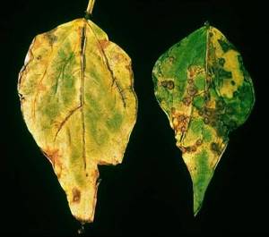
Common blight, fuscous blight (English)
|
Common blight, fuscous blight (photo: http://photos.eppo.org/index.php/image/ |
Scientific names
Xanthomonas axonopodis pv. phaseoli (Smith) Vauterin et al
Other scientific names
Xanthomonas campestris pv. Phaseoli (Smith) Dye Xanthomonas phaseoli var. fuscans (Burkholder) Starr & Burkholder
Significance
Important (spread and loss of yields).
Symptoms
On leaves: initially small water soaked lesions develop narrow, yellow halos. Lesions may enlarge and coalesce, causing extensive necrosis. Lesions may also occur on stems and pods. Infected seeds are sometimes wrinkled and the hilum may be discoloured. Symptoms similar to halo-blight of bean.
On seed: If the infection occurred when the pods were young, the seed may rot or be variously wrinkled and shrivelled. If the bacteria enter by way of the funiculus, only the hilum may be discoloured, but this is difficult to detect on dark-seeded varieties. Strains producing the brown pigment (so-called fuscans strains) give more conspicuous seed discoloration.
Hosts
The principal host is Phaseolus vulgaris and Phaseolus lunatus but other legume species are naturally infected, including P. lunatus, Vigna aconitifolia, V. radiata, and Vigna umbellate. Lablab purpureus and Mucuna deeringiana are possibly natural hosts. P. coccineus, P. acutifolius and Lupinus polyphyllus are hosts only by artificial inoculation (Bradbury, 1986). Macroptilium lathyroides.
Geographic distribution
EPPO region: Found in Egypt, Finland (unconfirmed), Lithuania, Moldova, Morocco (unconfirmed), Norway, Poland (unconfirmed), Sweden (unconfirmed). Widespread in Bulgaria, Hungary, Lebanon and Spain; locally established in France, Germany, Greece, Italy, Netherlands, Portugal (Madeira), Romania, Russia (European), Slovakia, Slovenia, Switzerland, Turkey and Yugoslavia. Found in the past but not established in the Czech Republic, Israel.
Asia: Bangladesh, Brunei Darussalam, Cambodia, China (Heilongjiang, Henan, Hunan, Jilin, Jiangsu, Liaoning, Zhejiang), Cyprus, Georgia, Hong Kong, India (Delhi, Maharashtra, Rajasthan, Uttar Pradesh), Indonesia, Israel, Japan, Lebanon, Korea Democratic People's Republic, Korea Republic, Malaysia, Myanmar (Burma), Nepal, Philippines, Sri Lanka, Taiwan, Thailand, Turkey, United Arab Emirates (IMI, 1996), Viet Nam, Yemen.
Africa: Angola, Burundi, Central African Republic, Egypt, Ethiopia, Kenya, Lesotho, Madagascar, Malawi, Mauritius, Morocco, Mozambique, Nigeria, Rwanda, Somalia, South Africa, Sudan, Swaziland, Tanzania, Tunisia, Uganda, Zaire, Zambia, Zimbabwe.
North America: Bermuda, Canada (Ontario), Mexico, USA (more prevalent east of the Rocky Mountains: Colorado, Hawaii, Michigan, Montana, Nebraska, New York, Texas, Wisconsin, Wyoming).
Central America and Caribbean: Widespread in Central America; Barbados, Costa Rica, Cuba, Dominica, Dominican Republic, El Salvador, Guatemala, Honduras, Jamaica, Martinique, Nicaragua, Panama, Puerto Rico, St. Vincent and Grenadines, Trinidad and Tobago.
South America: Argentina, Brazil (widespread), Chile, Colombia, Ecuador, Paraguay, Uruguay, Venezuela.
Oceania: Australia (New South Wales, Queensland, Victoria, Western Australia), New Zealand, Samoa.
EU: Present.
VeryVery widespread (Anonymous, 1971).
Biology and transmission
The bacterium enters the leaves via stomata or wounds, and subsequently invades the intercellular spaces, causing a gradual dissolution of the middle lamella. The stem is entered in three ways: via the stomata of the hypocotyl and epicotyl; through the vascular system of the leaf; or from infected cotyledons. The seed is penetrated via the vascular system of the pedicel and funiculus. The micropyle also serves as a point of entry into the seed. Direct penetration of seed has not been observed. The pathogen either remains in the seedcoat or passes to the cotyledons when the seed germinates, and so infection of the young plant results.
The bacterium can remain viable for several years beneath the seedcoat.
The disease is severe under conditions of high rainfall and humidity, with maximum development around 28°C. Dissemination in the field occurs in wind-driven rain, and insects.
Detection/indexing method
- At CIAT: Agar plate dilution technique. MXP (Claflin et al 1987) and YDCA semiselective culture medium and serology.
Treatment/control
- Procedure followed in case of positive test
References of protocols at EPPO, NAPPO or other similar organization
OEPP/EPPO Data sheets on quarantine organisms. Prepared by CABI and EPPO for the EU under Contract 90/399003
References and further reading
Anonymous. 1971. CMI distribution maps of plant diseases. No. 401 (edition 2).
Commonwealth Agricultural Bureaux, Slough.
Bradbury JF. (1986) Guide to plant pathogenic bacteria. CAB International, Wallingford, UK.
Claflin LE, Vidaver AK, Sasser M. (1987) MXP, a semiselective medium for Xanthomonas campestris pv. phaseoli. Phytopathology 77, 730-734.
Frison EA, Bos L, Hamilton RI, Mathur SB, Taylor JD. (eds.). 1990. FAO/IBPGR Technical Guidelines for the Safe Movement of Legume Germplasm. Food and Agriculture Organization of the United Nations, Rome/International Board for Plant Genetic Resources, Rome.
|
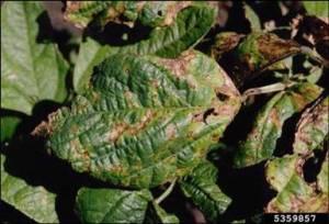
Photo by Howard F. Schwartz, Colorado State University, Bugwood.org |
Scientific names
Pseudomonas syringae pv. phaseolicola
Other scientific names
Pseudomonas savastanoi pv. phaseolicola
Significance
Important (spread and loss of yields).
Symptoms
Symptoms first appear as dark, water-soaked leaf spots, up to 3 mm in diameter and sometimes surrounded by a broad lemon-coloured halo. No lesions have been observed on stems or leaf stalks. When these pathogenic bacteria enter the leaves through wounds or natural openings like stomates they multiply rapidly and induce the formation of lesions. The lesions, which are watersoaked at first, soon become brown and dry and are usually surrounded by a yellow halo. When many lesions grow together, large areas of dead leaf tissue may develop. The halo is actually caused by a toxin that is produced by the bacteria within the lesion. As the toxin diffuses out into the leaf tissue it causes breakdown of chlorophyll and creates the halo. When toxin from infected leaves is translocated to the growing point of the shoot, the new leaves that develop are often stunted and chlorotic.
Hosts
Cajanus cajan, Lablab purpureus, Macroptilium spp., Phaseolus coccineus, P. lunatus, P.vulgaris, Pueraria spp., Vigna angularis, V. radiata, Neonotonia wightii. Isolates of the pathovar are categorised into three races on the basis of the reactions of a range of differential bean cultivars.
Geographic distribution
Worldwide (Anonymous, 1973). In temperate climatic conditions and in the tropics at medium to high altitudes (1000-2500 m). Race 3 of the pathogen has been found only in East and Central Africa.
Biology and transmission
Cell of the pathogen are single, straight rods and move by multitrichous polar flagellar. They are Gram-negative and strictly aerobic. The optimal growth temperature is 20 - 23o and on agar the bacterium produces white to cream coloured colonies wich exhibit a bluish tinge and often a green fluorescent pigment. P. syringae pv. phaseolicola survives in infected seeds and in vegetable(plant) residues in the surface of the soil until the environmental conditions are propitious for the development of the infection.
Detection/indexing method
- At CIAT: Agar plate dilution technique. King B semiselective culture medium and serology.
Treatment/control
- Procedure followed in case of positive test
References and further reading
Frison EA, Bos L, Hamilton RI, Mathur SB, Taylor JD. (eds.). 1990. FAO/IBPGR Technical Guidelines for the Safe Movement of Legume Germplasm. Food and Agriculture Organization of the United Nations, Rome/International Board for Plant Genetic Resources, Rome.
Lenné JM, Trutman P. 1990. Diseases of Tropical Pasture Plants. CAB Interantional in association with Natural Resource Institute and CIAT.
Lascano CE, Spain JM. Establecimiy Renovación de Pasturas. Red Interacional de Evaluación de Pastos Tropicales. Veracruz, Mexico 1988
Schwartz HF. 1989. Halo blight In: Schwartz, H.F. and Pastor Corrales, M.A. Bean Production Problems in the tropics. CIAT. Cali, Colombia, pp 285-302
Taylor JD. 1970. The quantitative estimation of the infection of bean seed with Pseudomonas phaseolicola (Burkh.) Dowson. Ann appl. Biol. 66: 29-36.
Bacterial blight, Bacterial Pod Rot
Scientific names
Pseudomonas fluorescens biotipo II Migula
Other scientific names
Bacillus fluorescens Trevisan 1889
Bacterium fluorescens (Trevisan 1889) Lehmann and Neumann 1896
Liquidomonas fluorescens (Trevisan 1889) Orla-Jensen 1909
Bacillus fluorescens Flgge 1886
Symptoms
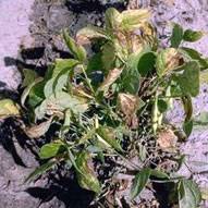
The disease is characterizates by water-soaked lesions on young growth especially young leaves, petioles and terminals, progressing to bliht, necrosis, dieback and defoliation. Necroticspots develop on older leaves and flowering and seed productions may be severely reduced.
Hosts
Centrosema sp. (C. acutifolium, C. pubescens, C. brasilianum, C. macrocarpum, C. schiedeanum and C. virginianum), Allium, Brassica, Phaseolus sp., Solanum spp., Leucaena leucocephala.
Geographic distribution
Colombia and Costa Rica.
Biology and transmission
Although the species is normally considered to be a saprophyte, Biotipe II causes root rots and leaf blights on a range of hostd, including legumes. It is distinguished from other fluorescent pseudomonas by the following characterisitic: more than one polar flagellum, no poyocyanin or carotenoid pigments produced, no growth at 41o C, gelatin hydrolysed but not starch and utilization of a wide range of carbon sources. Disease is favoured by high relative humidity and moderately high temperatures. The bacterium can survive unfavourable periods on affected plants and soil for as long as 6 weeks. The bacterium is seed-borne and levels of infections as high as 32 % have been found in seed lots of C. acutifolium.
Detection/indexing method
- At CIAT: Agar plate dilution technique. King B semiselective culture medium.
Treatment/control
- Procedure followed in case of positive test
References and further reading
Arias B, Lenne JM. 1988. Sistemas de produccion de Pasturas Tropicales de semillas de Centrosema acutifolium y efecto de bacterioocidas en la incidencia de Pseudomonas fluorescens Biotipo II. Pasturas Tropicales Vol. 10, 11-18.
Bradbury JF. 1986. Guide to plant pathogenic Bacteria, CAB Interantional, Wallingford, UK
Guevara Gómez CL, Lenné JM, Torres GC. 1983. Etiology of dieback of Centrosema spp. and effect of the pathogen on yield and quality during the period of establishment of the legume. Phytopathology 73, 122
Lenné JM, Torres GC, Victoria JI. 1981. Bacterial leaf spot and dieback of Centrosema spp. Proc. Fifth Int. Conf. Plant Path. Bact Cali, 35-38.
 |
 |
Scientific names
Curtobacterium flaccumfasciens pv. flaccumfaciens (Hedges) Collins & Jones
Other names
Bacterial wilt (Phaseolus beans), bacterial tan spot (soyabeans)
Symptoms
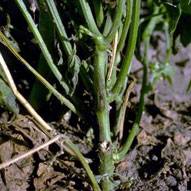
Seedlings are stunted, wilted and usually die. In some plants the affected parts are dull green in colour and sometimes breaking of the stems. Infected pods show discoloured sutures and may show yellowish areas.
Foliar interveinal chlorosis and necrosis. When the lower stem and root are cut longitudinally and observed, the vascular system is often discolored brown to black. The younger the plant becomes infected, the more severe the damage to the plant. Seedlings are frequently severely stunted or killed. If plants survive to maturity, seeds may show yellow or purple discoloration.
Seeds of white-seeded cultivars, when infected systemically, are bright-yellow; in cultivars with coloured seed coats, the coloration is less conspicuous. There may be a little yellow slime at the hilum, and seeds may be variously shrivelled. The colour mutants, aurantiacum and violaceum, produce an orange and purple discoloration, respectively, in the seedcoat.
Hosts
Lablab purpureus, Phaseolus coccineus, Phaseolus lunatus, Phaseolus vulgaris, Vigna angularis, Vigna unguiculata, Zornia spp. and possibly Glycine max.
All members of Leguminosae.
Geographic distribution
Bacterial wilt of beans has been reported in North and South America, Europe, and Australia
EPPO region: Recorded in Albania, Ukraine. Found but not established in Greece and Hungary; locally established in Bulgaria (unconfirmed), Romania, Tunisia, Turkey (unconfirmed), Russia (Far East, Southern Russia; only on soyabean) and Yugoslavia. Reports from Belgium (OEPP/EPPO, 1982), France, Germany and Switzerland have not been substantiated.
Asia: Russia (Far East), Turkey (unconfirmed).
Africa: Mauritius, Tunisia.
North America: Canada (Ontario), Mexico (unconfirmed), USA (first reported in 1920, especially in irrigated high plains and Midwest, but not reported since early 1970s except in Iowa on soyabeans; Hall, 1991. Specific records from Colorado, Connecticut, Iowa, Idaho, Michigan, Montana, Nebraska, Ohio, Oregon, Virginia, Wisconsin).
South America: Colombia, Venezuela.
Oceania: Australia (New South Wales, Queensland, South Australia, Victoria).
Europe: Present (Belgium, Bulgaria, Greece, Hungary, Romania, Yugoslavia.
Biology and transmission
Seed transmitted externally or internally in Phaseolus vulgaris and possibly in Glycine max. The pathogen can survive from 5 to 24 years in seeds.
Seed is the most important means of survival and spread. The bacteria can overwinter on weeds or in crop debris in the field. In the field it has been known to survive in soil for at least two winters between bean crops rotated with wheat. There are no reports of vectors, but the nematode Meloidogyne incognita may assist entry by providing wounds.
C. flaccumfaciens pv. flaccumfaciens can infect in the absence of rain; it has not been observed to enter via stomata. Once within the plant, the bacterium colonizes the vascular tissue.
Detection/indexing method
- At CIAT: Agar plate dilution technique. NBY (Nutrient Broth Yeast extract) semiselective culture medium and serology.
Treatment/control
- Procedure followed at the centers in case of positive test
References of protocols at EPPO, NAPPO or other similar organization
OEPP/EPPO (1982) Data sheets on quarantine organisms No. 48, Corynebacterium flaccumfaciens. Bulletin OEPP/EPPO Bulletin 12 (1).
References and further reading
Frison EA, Bos L, Hamilton RI, Mathur SB, Taylor JD. (eds.). 1990. FAO/IBPGR Technical Guidelines for the Safe Movement of Legume Germplasm. Food and Agriculture Organization of the United Nations, Rome/International Board for Plant Genetic Resources, Rome.
Hedges F. (1926) Bacterial wilt of beans (Bacterium flaccumfaciens Hedges), including comparisons with Bacterium phaseoli. Phytopathology 16, 1-22.
Torres GC, Lenné JM, Victoria JI. Bacterial wilt of Zornia spp. caused by Corynebacterium flaccumfaciens. Conference paper Proceedings of the Fifth International Conference on Plant Pathogenic Bacteria. 1982 pp. 74-79.